HORMONE MEASUREMENT GUIDELINES: Tracing lipid metabolism: the value of stable isotopes
-
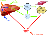 Figure 1
Sites of utilisation of palmitate, glycerol, water and acetate tracers used for the measurement of lipid metabolism.
-
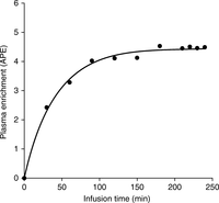 Figure 2
Schematic example of a constant infusion of tracer to achieve an isotopic steady state.
-
Figure 3
Calculating rate of appearance (Ra) at isotopic steady state. Rd, rate of disappearance; black circles represent the tracer; white circles represent the tracee. Reproduced, with permission, from Wolfe RR & Chinkes DL (2004) Isotope Tracers in Metabolic Research: Principles and Practice of Kinetic Analysis, 2nd Edition. Copyright 2004 Wiley-Blackwell.
-
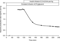 Figure 4
Glycerol production rate in a healthy subject measured with a constant infusion of [2H5]glycerol during fasting and following an infusion of insulin. The decrease in glycerol production rate is a measure of the insulin sensitivity of lipolysis.
-
Figure 5
Plasma NEFA oxidation calculated using the acetate correction factor when infusing [U-13C]palmitate. The correction factor is the fractional 13C-label recovery in breath CO2, observed after an infusion of [1,2-13C]acetate. Reproduced, with permission, from Schrauwen P, van Aggel-Leijssen DP, van Marken Lichtenbelt WD, van Baak MA, Gijsen AP, Wagenmakers AJ (1998) Validation of the [1,2-13C]acetate recovery factor for correction of [U-13C]palmitate oxidation rates in humans. Journal of Physiology 513 215–223. Copyright 2004, John Wiley and Sons.
-
Figure 6
Typical curve fit for leucine tracer:tracee ratio in VLDL, IDL and LDL apoB100 using mathematical modelling following an infusion of [1-13C]leucine. VLDL shown as large squares. IDL shown as triangles. LDL shown as circles. The solid lines indicate the curve fit. The modelling provides a measure of VLDL, IDL and LDL apoB100 fractional catabolic rate and production rate.
-
Figure 7
Typical curve fit of VLDL1 and VLDL2 glycerol tracer:tracee ratio in TAG, following an i.v. bolus of [2H5]glycerol, using mathematical modelling. VLDL1 shown as large squares. VLDL2 shown as small circles. The solid lines indicate the curve fit. The modelling provides a measure of VLDL1 and VLDL2 TAG fractional catabolic rate and production rate. Reproduced, with permission, from Sarac I, Backhouse K, Shojaee-Moradie F, Stolinski M, Robertson MD, Bell JD, Thomas EL, Hovorka R, Wright J & Umpleby AM (2012) Gender differences in VLDL1 and VLDL2 triglyceride kinetics and fatty acid kinetics in obese postmenopausal women and obese men. Journal of Clinical Endocrinology and Metabolism 97 2475–2481. Copyright 2012 The Endocrine Society.
-
Figure 8
Glycerol tracer:tracee ratio in chylomicron TAG in healthy subjects (white circles) and subjects with MetS (black circles) following an i.v. bolus of [2H5]glycerol (mean±s.e.m.). Mathematical modelling of this data provides a measure of chylomicron TAG fractional catabolic rate and production rate. Reproduced, with permission, from Shojaee-Moradie et al. (2013). Diabetes, American Diabetes Association, 2013. Copyright and all rights reserved. Material from this publication has been used with the permission of American Diabetes Association.
- © 2015 Society for Endocrinology