Cortisol mobilizes mineral stores from vertebral skeleton in the European eel: an ancestral origin for glucocorticoid-induced osteoporosis?
- Miskal Sbaihi1,
- Karine Rousseau1,
- Sylvie Baloche1,
- François Meunier1,
- Martine Fouchereau-Peron1,2 and
- Sylvie Dufour1
- 1Muséum National d'Histoire Naturelle, DMPA USM 0401, UMR 7208 CNRS BOREA ‘Biologie des Organismes et Ecosystèmes Aquatiques’, 7 rue Cuvier, CP 32, 75231 Paris Cedex 05, France2Marine Station of Concarneau, 29182 Concarneau Cedex, France
- (Correspondence should be addressed to S Dufour; Email: dufour{at}mnhn.fr)
-
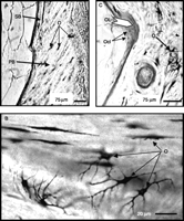 Figure 1
Histology of eel vertebral bone. Vertebrae were stained with basic fuchsine, embedded in methacrylate and cut at 30±5 μm (A) Vertebral section from a control eel showing primary bone (PB) and secondary bone (SB) indicating bone remodeling. Many osteocytes (O) are included in the bone structure demonstrating a cellular (osteocytic) bone (Scale bar=75 μm). (B) Higher magnification of a vertebral section from a control eel showing the spangled form of the osteocytes (O; Scale bar=20 μm). (C) Vertebral section of a cortisol-treated eel showing a large osteoclastic lacuna (OL) and the presence of osteoclasts (Ocl; Scale bar=75 μm).
-
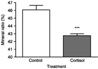 Figure 2
Effect of cortisol on mineral ratio (MR) of eel vertebrae. MR is measured by incineration. MR=(mineral weight/dry weight)×100. Results are expressed as mean±s.e.m. (n=8 eels/group). ***P<0.001 as compared with controls.
-
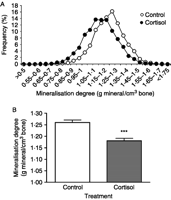 Figure 3
Effect of cortisol on mineralization degree (MD) of eel vertebrae. MD (g mineral/cm3 of bone) is measured by quantitative microradiography of vertebral sections (100±1 μm) of control and cortisol-treated eels. (A) Distribution of MD values in vertebrae from control and cortisol-treated eels (eight vertebral zones measured per eel vertebral section; eight control eels and eight treated eels). (B) Mean MD±s.e.m. of vertebrae from control and cortisol-treated eels (n=8 eels/group). ***P<0.001 as compared with controls.
-
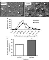 Figure 4
Effect of cortisol on periosteocytic resorption of eel vertebrae. (A) Microradiographs of ultrathin vertebral sections (<20 μm) from control and cortisol-treated eels, showing periosteocytic lacunae (arrows). The surface areas of lacunae are measured by image analysis of microradiographies. Osteocytic lacunae are larger in cortisol-treated eels than in controls. (Scale bar=10 μm). (B) Distribution of surface area of periosteocytic lacunae in vertebral sections of control and cortisol-treated eels (40 osteocytic lacunae measured/eel; eight control eels and eight treated eels). (C) Mean surface area of periosteocytic lacunae in vertebrae from control and cortisol-treated eels. Results are expressed as mean±s.e.m. (n=8 eels/group). ***P<0.001 as compared with controls.
-
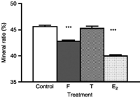 Figure 5
Comparison of the effects of cortisol and sex steroids on mineral ratio. MR is measured by incineration. MR=(mineral weight/dry weight)×100. Results are expressed as mean±s.e.m. (n=8 eels/group). ***P<0.001 as compared with controls.
-
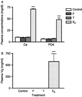 Figure 6
Comparison of the effects of cortisol and sex steroids on plasma concentrations of calcium, phosphate (A) and vitellogenin (B) in the European eel. Plasma concentrations of calcium and phosphate are measured spectrophotometrically and vitellogenin by homologous ELISA. Results are expressed as mean±s.e.m. (n=8 eels/group). ***P<0.001 as compared with controls.
- © 2009 Society for Endocrinology


