15 YEARS OF PARAGANGLIOMA: Imaging and imaging-based treatment of pheochromocytoma and paraganglioma
- Department of Endocrinology, La Timone University Hospital, Aix‐Marseille University, Marseille, France
1Department of Biophysics and Nuclear Medicine, Medical University Innsbruck, Innsbruck, Austria
2Diagnostic Nuclear Medicine Division, Department of Nuclear Medicine, All India Institute of Medical Sciences, New Delhi, India
3Program in Reproductive and Adult Endocrinology, <ce:italic>Eunice Kennedy Shriver</ce:italic> National Institute of Child Health and Human Development, National Institutes of Health, Bethesda, Maryland, USA
4Department of Nuclear Medicine, European Center for Research in Medical Imaging (CERIMED), La Timone University Hospital, Aix‐Marseille University, 264, rue Saint‐Pierre, 13385 Marseille, France
5Institut Paoli‐Calmettes, Inserm UMR1068 Marseille Cancerology Research Center, Marseille, France
- (Correspondence should be addressed to D Taieb; Email: david.taieb{at}ap-hm.fr)
-
Figure 1
4D MR angiography in a left tympanic PGL. (A) Selected dynamic images showing early arterial enhancement of the PGL (arrows). (B) Volumetric interpolated fat-saturated (FATSAT) T1-weighted (VIBE) (left), an informative image extracted from 4D MR angiography (middle), fusion image with a CT scan for better evaluation of temporal bone extension (right).
-
Figure 2
Head-to-head comparison of 18F-FDOPA PET/CT, 68Ga-DOTATATE PET/CT, CT, T1 SE Gado MR, and angiography MR sequences in a multifocal SDHD-related PGL. Axial anatomical and functional images centered over the jugular foramen: red arrow: right JP, black arrow: left JP. Note that the small right JP (red arrow) is missed by angio CT and T1 SE Gado sequence due to the gadolinium enhancement of the right internal jugular vein. By contrast, early arterial images on 4D MR angiography or TOF angiography images have detected this tiny PGL (red arrow). In the present case, the MR imaging protocol was adjusted according to the functional imaging findings. Note the higher tumor uptake with 68Ga-DOTATATE compared to 18F-FDOPA.
-
Figure 4
Functional imaging findings in a case of adrenal lymphoma and a case of metastatic pheochromocytoma. (A) Adrenal lymphoma with a mediastinal lymph node (arrows). (B) Sporadic pheochromocytoma with a metastatic mediastinal lymph node (arrows). This patient's tumors have rapidly metastasized to the lungs. Note the similar 18F-FDG PET/CT presentation in both cases. As expected, the PHEO was positive for 18F-FDOPA.
-
Figure 5
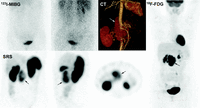
Functional imaging findings in a case of extraadrenal sympathetic SDHB-related PGL. Multimodality imaging with 123I-MIBG (anterior and posterior views), SRS (three images), and 18F-FDG PET/CT. The PGL (arrow) was moderately avid for Octreoscan and quasi-negative on MIBG scan. In contrast, the tumor exhibited high 18F-FDG uptake, a finding that is typical for SDHB-tumors.
- © 2015 Society for Endocrinology


