The diagnosis and management of parasellar tumours of the pituitary

  1. Ashley B Grossman3
  1. Endocrine Unit, Department of Pathophysiology, National University of Athens, Athens, Greece1Department of Academic Radiology, St Bartholomew's Hospital, London, UK2Department of Endocrinology, Theagenion Hospital, Thessaloniki, Greece3Department of Endocrinology, St Bartholomew's Hospital, London EC1A 7BE, UK
  1. (Correspondence should be addressed to A B Grossman; Email: a.b.grossman{at}qmul.ac.uk)
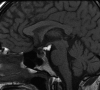
  1. Figure 1

    Sagittal enhanced T1-weighted image of a hypothalamic glioma. This is partially cystic, partially solid and involves the chiasm and the hypothalamus.

  2. Figure 2

    Sagittal enhanced T1-weighted image of a germinoma with both a suprasellar and pineal enhancing mass lesions.

  3. Figure 3

    Coronal enhanced T1-weighted image of lymphoma. There is abnormal dural enhancement of both cavernous sinuses extending to involve the pituitary gland itself and with slight thickening of the stalk.

  4. Figure 4

    Sagittal unenhanced T1-weighted image of a craniopharyngioma. The mass is inseparable from the chiasm and hypothalamus and is predominantly of low T1 signal suggesting a cystic mass, although without a high lipid content.

  5. Figure 5

    Sagittal enhanced T1-weighted image of a solidly enhancing craniopharyngioma. This mass is inseparable from the hypothalamus and chiasm. There is an area of low signal within the mass representing calcification (confirmed on CT).

  6. Figure 6

    Coronal enhanced T1-weighted image of a chondrosarcoma. This is an enhancing mass involving the left side of the sella and the cavernous sinus, but arising from the skull base just off midline.

  7. Figure 7

    Coronal enhanced T1-weighted image of Langerhans' cell histiocytosis. There is diffuse enlargement and enhancement of both cavernous sinuses. The pituitary gland is bulky, although the stalk is not thickened.

  8. Figure 8

    Sagittal enhanced T1-weighted image of a suprasellar meningioma. The mass lies above the pituitary (which is normal), involves the chiasm and extends anteriorly along the planum sphenoidale.

  9. Figure 9

    Coronal enhanced T1-weighted image of a meningioma in the left cavernous sinus. The meningioma enhances avidly, expands the cavernous sinus and is causing some narrowing of the left cavernous carotid artery, which is of smaller calibre than the right carotid.

  10. Figure 10

    Sagittal enhanced T1-weighted image of a Rathke's cleft cyst. The cyst is seen to be non-enhancing and lying on the superior aspect of the pituitary gland that appears flattened. It is causing elevation of the optic chiasm.

  11. Figure 11

    Sagittal enhanced T1-weighted image of a hypothalamic hamartoma. This is seen as a pedunculated mass hanging from the undersurface of the hypothalamus. It does not enhance and has the same signal as the grey matter of the brain parenchyma.

  12. Figure 12

    Sagittal unenhanced T1-weighted image of a suprasellar lipoma. The high signal mass containing fat is seen lying on the undersurface of the hypothalamus in the interpeduncular cistern.

  13. Figure 13

    Coronal enhanced T1-weighted image of a left sided Schwannoma of the trigeminal nerve. There is an enhancing mass expanding the left cavernous sinus which followed the course of the trigeminal nerve on other images.

  14. Figure 14

    Sagittal enhanced T1-weighted image of a granular cell tumour. This enhancing mass lesion involves the pituitary stalk and the median eminence.

| Table of Contents