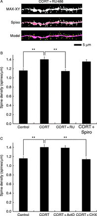
Effects by the blockade of receptors and inhibition of gene expressions on CORT-induced spinogenesis. (A) Representative images of confocal micrographs; the spines along dendrite with 30 nM CORT and 10 μM RU486 (blocker of glucocorticoid receptor (GR)) (CORT+RU) for 1 h. Maximal intensity projections onto XY plane from z-series confocal micrographs (MAX-XY), images analyzed by Spiso-3D (Spiso) and three-dimensional model illustrations (model) are shown together. Bar, 5 μm. (B) Effects from the blocker of GR and mineralocorticoid receptor (MR) on the CORT-induced spinogenesis. A 1 h treatment in ACSF without drugs (control), with 30 nM CORT, with 30 nM CORT and 10 μM RU486 (CORT+RU), and with 30 nM CORT and 10 μM spironolactone (blocker of MR) (CORT+Spiro). Vertical axis represents the average number of spines per 1 μm of dendrite. Data are represented as mean±s.e.m. (C) Effects by the inhibition in mRNA and protein synthesis on the CORT-induced spinogenesis. A 1 h treatment in ACSF without drugs (control), with 30 nM CORT, with 30 nM CORT and 4 μM actinomycin D (translation inhibitor) (CORT+ActD), and with 30 nM CORT and 20 μM cycloheximide (transcription inhibitor) (CORT+CHX). Vertical axis represents the average number of spines per 1 μm of dendrite. Data are represented as mean±s.e.m. The statistical significance yielded: **P<0.01 vs ‘CORT’. For each drug treatment, we investigated three to four rats, six to eight slices, 30–40 neurons, 60–80 dendrites, and ∼3000–4000 spines. A full colour version of this figure is available at http://dx.doi.org/10.1530/JOE-15-0078.